
Whole-genome and exome sequencing, delivered as a short list of variants that matter.
We read a whole genome or all its protein-coding genes, find where it differs from the reference and classify the variants that could matter for your question.
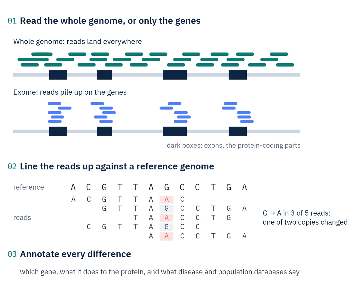
Line the reads up, then look for differences.
Whole-genome sequencing reads the entire genome: genes, the stretches between them and the regulatory regions. Exome sequencing first pulls out the exons, the protein-coding parts that make up about 1.5% of the human genome, and reads only those. The exome costs less per sample; the genome also sees non-coding and regulatory variants.
The short reads are placed on a reference genome. Where many reads show a different letter at the same position, the sample carries a variant. Coverage, the number of reads stacked on each position, decides how confidently a variant is called: about 30 reads for a human genome, more for an exome.
One human exome yields well over a hundred thousand variants, and almost all of them are harmless. We keep the variants that independent calling methods agree on, set aside those that are common in the population, and classify the rest on the five-tier ACMG scale from pathogenic to benign.
Key terms
- Exome
- All protein-coding parts of the genome, about 1.5% of human DNA.
- Coverage
- How many reads, on average, cover each position. 30× means 30 reads.
- SNV and indel
- A single-letter change, and a small insertion or deletion of letters.
- Heterozygous
- Only one of the two chromosome copies carries the variant, so about half the reads show it.
- Population frequency
- How common a variant is in large reference populations. Common variants rarely cause rare disease.
- ACMG classification
- The five-tier scale for grading variants: pathogenic, likely pathogenic, uncertain, likely benign, benign.
Bring the question. We design the experiment around it.
- Which variant in this genome could explain the phenotype?
- Is this variant rare, and is it likely to damage the protein?
- How should each candidate variant be classified?
- Do I need the whole genome, or is the exome enough?
Where it is used
- Rare and inherited disease researchSearching a genome or exome for the variant behind a phenotype.
- Candidate gene screeningChecking the genes linked to a condition for rare, damaging variants.
- Viral genomesConsensus genomes, variants and family trees of viruses.
- Bacterial isolatesWhole genomes of isolates for characterisation.
- Plant and animal genomesResequencing of lines, breeds and strains against their reference.
- Species without a referenceWhen no reference exists, the genome is assembled first.
Four steps from DNA to a classified variant list.
- Step 1
Choose genome or exome
We match the method and coverage to the question: exome for coding variants, genome when non-coding regions matter too.
- Step 2
Check the DNA
DNA amount and integrity are checked before library preparation, so problems show up early.
- Step 3
Sequence
Libraries are sequenced as 150-letter read pairs to the agreed coverage.
- Step 4
Call, filter and classify
Variants from several callers are compared, filtered by population frequency and predicted impact, classified and summarised in an interactive report.
What the analysis shows you.
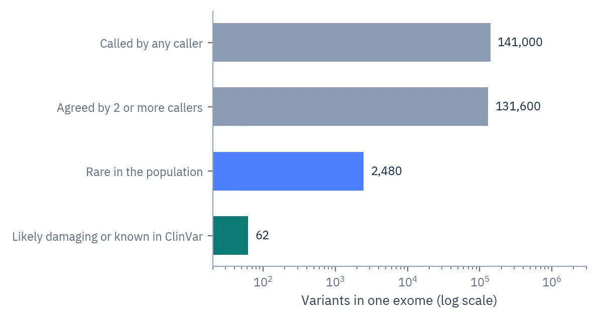
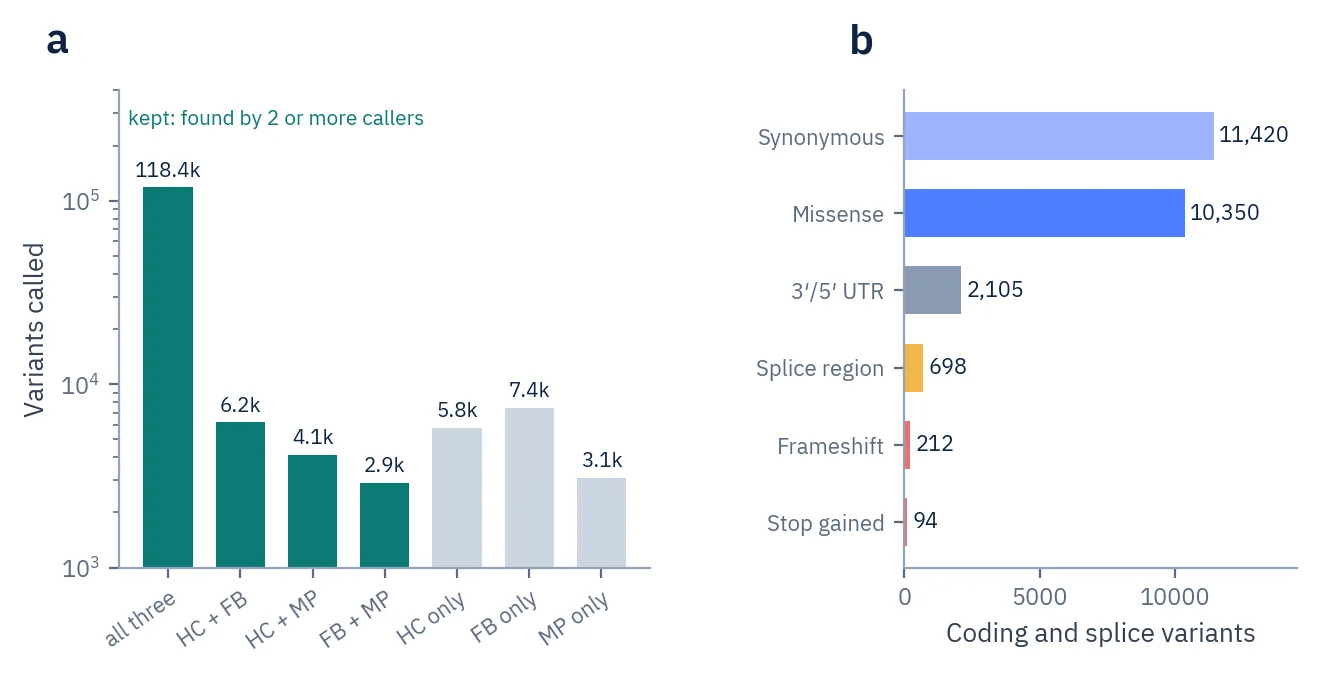
From raw calls to a shortlist
Each filter removes variants that are common or unlikely to matter. Of about 140,000 calls in one exome, a few dozen are left to classify.
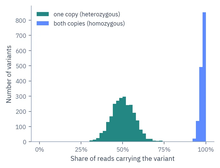
One copy or both?
The share of reads carrying each variant. Variants on one chromosome copy sit near 50%, variants on both copies near 100%.
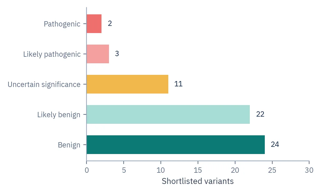
Classified variants
The shortlist graded on the five-tier ACMG scale, with the reasoning behind each classification in the report.
Included as standard every project
- Alignment and clean-upReads are placed on the reference genome, with duplicates marked and base qualities recalibrated.
- Consensus variant callingSeveral independent callers are run and the variants most of them agree on are kept. More run in parallel where the project needs it.
- AnnotationEach variant is linked to its gene, its effect on the protein, and population and disease databases.
- FilteringRare variants that are predicted to be damaging or known as pathogenic are shortlisted.
- Classification and reportACMG classification with reasoning and literature, in an interactive report with a list of critical findings.
Added for your question custom
- Viral genomesConsensus genomes, variants and phylogenetic trees from viral sequencing data.
- Somatic tumour reportsTumour variants reported following ClinGen, CGC and VICC guidelines.
- HLA typing and copy numberHLA alleles, and gains or losses of DNA segments.
- Genome size checkFor less-studied organisms, genome size is looked up in public databases before the depth is set.
What goes in, and how we run it.
- Sequencing
- Illumina, 150 bp read pairs
- Typical coverage
- About 30× for a human genome · 50–100× for germline exomes · 100× or more for tumour exomes
- Variants
- Single-letter changes (SNVs) and small insertions and deletions
- Samples
- Blood, tissue and extracted DNA · bacterial isolates, plants and animals
- Delivered
- Consensus VCF, annotated and classified variant tables, interactive report
- Main tools
- nf-core/sarek, BWA-MEM, HaplotypeCaller, FreeBayes, OpenCRAVAT
Figures, interpretation and Methods, ready for the manuscript.
- ReportInterpretation written against your hypothesis
- FiguresPublication-ready figures for the manuscript
- MethodsMaterials & Methods text for the paper
- Tables and dataAll results as tables, plus the processed data files
Genome or exome?
Bring the question to the call and the scientist recommends one, with the reasoning for your samples.
| Aspect | Whole genome | Whole exome |
|---|---|---|
| Covers | Every part of the genome, including non-coding regions | Protein-coding exons only |
| Usual depth | About 30× for a human genome, and deeper when the question needs it | Usually 50× to 100×, and far deeper for tumour or low-frequency work |
| Data per sample | Reads spread across 3 billion letters | Far less data in total, concentrated on the exons |
| Finds | Coding and non-coding variants | Coding variants |
| Best when | Regulatory or non-coding variants matter, or an exome found nothing | Many samples, with coding variants as the main suspects |
Straight answers before you email.
Related services
Which organisms can you sequence?
Human, other mammals, plants, insects, fish, bacteria, fungi, viruses, cell lines, etc. Ask about yours. The lab work is the same for any DNA. What changes is the interpretation. Human variants have decades of databases behind them. Most other species have very little, so the answer comes from comparing your own samples: your lines against each other, your isolates against a collection, your clone against its parent, etc. On the call we check what reference and databases exist for your species first, then design the comparison around what is actually there.
Whole genome or exome, how do I choose?
The exome is the protein-coding part, plus the splice sites next to it. It needs far less sequencing per sample, and most known disease-causing variants sit inside it. Good capture kits exist for human and a few model organisms only, so for other species it is usually a whole genome or a custom panel. Go for the whole genome if you care about non-coding or regulatory variants, or about large deletions and duplications. Bring the question to the call and the scientist tells you which one actually answers it.
What does exome sequencing miss?
Everything outside the targeted regions, which is most of the genome. Also parts of the targets themselves, because capture is uneven. Regions rich in G and C, repeats, duplicated genes, etc. often come out with too few reads. So a gene with no variants is not the same as a gene that was read properly. We send you the coverage for every gene on your list. And if your key genes sit in a weak region of a kit, the scientist tells you on the call, while you can still pick a different kit or move to a whole genome.
How much sequencing coverage do I need?
Depends what you are looking for. A variant you inherited sits on one of your two chromosome copies, so it turns up in about half the reads at that spot, and moderate depth is enough. A variant that is in some cells and not others, like in a tumour sample or a mixed culture, turns up in a small share of reads, so it needs much more. This is one of the things the call is for. You describe the question and the samples, and the scientist designs the depth around them.
How is sequencing coverage worked out?
Read length times number of reads, divided by the size of your genome or target region. That is the figure on paper. The real one is lower, because duplicate reads and reads that fail to map count for nothing. Coverage is also patchy, so a healthy average can still hide genes with almost no reads on them. We report the depth gene by gene, not just the average, and go through it with you before any interpretation starts.
How deep can you sequence?
There is no fixed ceiling. Depth is a design choice, so if your question needs more we run more. For tumour samples we often go to 500× or 1000×. In a very mixed tumour the mutation behind a neoantigen can sit in a small fraction of the cells, and at ordinary depth it is buried in the noise, so deep sequencing is what rescues it. The same applies to mosaic tissue, mixed cultures, low-frequency variants, etc. The real limit is not the sequencer but your sample, because once every unique DNA molecule in the library has been read, extra reads are only copies of ones you already have. Tell the scientist what share of cells you need to detect and the depth is set from there.
How big is my organism’s genome, and how much sequencing does that mean?
We check it before setting the depth, because coverage is sequencing divided by genome size. On one project two genomes turned out three times bigger than the published estimates. At the planned amount of sequencing both would have been far too thin to use. For less studied species the published size is often wrong or missing, and we can estimate it from the first data. The check is part of designing your experiment.
My cell line has been in culture a long time. Will that show in the data?
Often yes. Lines pick up mutations and copy number changes every time you passage them, and the fastest-growing cells take over. The same named line in two labs can differ across hundreds of genes and behave differently in experiments. Sequencing shows what your current culture actually carries. If you still have an early frozen vial, mention it on the call, because sequencing both gives you a direct comparison, and that tells you far more than one sample on its own.
My cell line came from a tumour, so there is no matched normal sample. What then?
Normal for tumour lines, and we can work with it. Without a normal sample we separate acquired from inherited variants using population databases, a panel of unrelated normals, modelling of read shares against tumour purity and copy number, etc. That gives likelihoods, not a clean split, so some calls come out solid and some do not. The report marks which are which, so you know what to confirm before you build on it. If any normal material from the same donor exists, even an old sample, say so on the call, because it changes the whole analysis.
How deep should a tumour sample be sequenced?
Deeper than inherited disease work, and how much deeper depends on the sample. The share of reads carrying a tumour mutation is not simply the fraction of tumour cells. It also depends on the copy number at that spot and on whether the mutation is in every tumour cell or only part of them. Low tumour content and part-of-the-tumour mutations both push that share down towards the error rate of the whole process, which comes from handling and library prep as much as from sequencing. Below a certain share, extra depth stops helping and the library has to be built with molecular tags so errors can be filtered out. Get the tumour content from your pathologist before the call, because it drives this decision.
What do I get from sequencing a bacterial isolate?
Species ID, typing so you can compare your isolates with each other and with public collections, resistance genes, virulence genes, plasmid content as far as short reads can reconstruct it, etc. The same data screens for contamination, though a clean screen only means nothing showed up above the detection limit. Tell the scientist which of these you need and they go into the design. If you need the chromosome and plasmids closed rather than in pieces, that takes long reads, which we also run.
Can you sequence viral genomes?
Yes. First question is whether your virus is DNA or RNA, because RNA needs a reverse transcription step and a different workflow. You get a consensus genome per sample, the variants against a reference, a tree showing how your samples relate to each other and to published sequences, etc. Minority variants inside the viral population can be reported when there is enough material and depth to tell them apart from errors, which is not true of every sample. Tell the scientist the sample type and how much virus you expect, because that decides whether we enrich for it.
How do you call variants?
Several callers, not one. They are not truly independent, because they read the same alignments and share some blind spots, but they handle difficult regions differently, so keeping what most of them agree on removes a lot of calls that are really one program’s quirk. It is a trade, not a free win: the stricter you are, the more real variants you lose along with the false ones. Where a project needs more callers we run more in parallel. Large rearrangements, variants present in only a few cells, etc. need their own specialised callers. Which ones and how many comes out of what your project is trying to show.
Will you find large deletions and duplications?
Usually yes. Short reads give three clues: read depth drops or rises across the region, read pairs map further apart than they should, and some reads sit right across the join and show it directly. The deletion does not have to fit inside one read. What makes it hard is repeats at the break points, where reads cannot be placed confidently. Exomes are weaker at this than whole genomes because they only sample the targets. If the rearrangement is the point of your study, say so on the call, because long reads pin down break points much better and we run those too.
Are there genes that short reads struggle with?
Yes. Genes with a near-identical twin elsewhere in the genome, genes inside duplicated stretches, etc., because reads cannot be assigned confidently between the copies. The same happens in other species wherever gene families are large or the genome has been duplicated. Many of these spots are already catalogued, so send your gene list before the design is set and we tell you which of your genes short reads cover properly and which they do not.
Which reference genome do you use, and does it matter?
It matters more than people expect, so it goes in the Methods with the version. For human work GRCh38 is the usual choice and most annotation databases are built on it. The complete T2T-CHM13 assembly covers regions GRCh38 leaves out and maps reads better in many of them. It is not a straight upgrade, because annotation databases are still catching up and some of the newly added repeat sequence still cannot be mapped with short reads. For other species the question is whether a better assembly has appeared since the last study on your system. We check that while designing your experiment and tell you which build we use and why.
How do you decide which variants matter?
Depends on the question. For an inherited disease we filter on how common a variant is in population databases, with the cut-off worked out from how common the disease is and how it is inherited, then shortlist what is rare and either predicted to break the protein or already published as disease-causing. Splice-site and regulatory variants are searched separately, because protein predictors miss them. Cancer, population traits, microbial work, plant breeding, etc. each need completely different filtering. We agree the strategy with you before running it, and you get the unfiltered data too, so anything we decided can be undone.
The variant I care about has never been described before. Can you still say anything about it?
Yes, quite a lot, though the honest start is that most rare variants turn out not to be the cause, and some stay unresolved however hard you work them.
Here is what we can use. Whether it is absent from population databases, which is weak evidence on its own and only counts if that spot is well covered there. How badly the gene tolerates damage, judged from large population data. Where it sits: a known functional part of the protein, a key residue, a position conserved across distant species, etc. What the change is predicted to do, scored with one predictor chosen in advance, because picking the tool that gave you the answer you wanted is not evidence. Whether it affects splicing, predicted separately, since many variants that look harmless in the protein still create or destroy a splice site. Whether the same change in a closely related gene is known to cause disease. And whether the mechanism fits, because a predicted truncation means a lot in a gene where loss of function causes the disease and little where it does not.
We switch on the annotation sources that apply to your genes and your organism, and the report says which lines of evidence existed for each variant and which did not. If one variant is the whole point of the study, tell the scientist and we work it up on its own.
How far do you go with ACMG classification?
Two levels, and you decide with the scientist which one your study needs. The standard level gives you the five-tier grade with the evidence behind each call. The detailed level works through the criteria one by one for a named variant: frequency against a threshold set for that gene and disease, computer prediction at calibrated strength, published lab experiments judged on whether the assay is actually relevant, inheritance in the family if you have the samples, whether the variant arose new, whether the mechanism fits the gene, etc. Where a ClinGen expert panel has published rules for your gene we use those instead of the general ones.
What happens when a variant comes back as uncertain?
It is reported as uncertain with the evidence laid out, because that is what the data supports. Uncertain means the evidence has not reached either threshold yet, not that the variant is suspicious, and these are very common. Where there is a real way to resolve it, the report names it: testing family members, RNA evidence, a lab assay, waiting for someone else to publish the same variant, etc. Where your material cannot settle it, the report says so. We then go through the uncertain calls with you and say which ones are worth chasing and what each one would take.
Can RNA data help resolve an uncertain variant?
Often, and for a variant predicted to hit splicing it is one of the strongest things you can add, because it replaces a prediction with an observation. Conditions apply. The gene has to be expressed in a tissue you can get, at enough depth to see the transcript. Faulty transcripts can also be destroyed by the cell before you see them, which makes a real effect look like nothing. Where you cannot get the right tissue, a validated lab assay can sometimes supply the evidence instead. We run RNA sequencing, so it can be designed into the same project.
Can a variant classification change later?
Yes. A classification reflects the evidence that existed on the day. Among uncertain variants that do get reclassified, most studies find they move towards benign. How often reclassification happens at all varies a lot by gene and by how long you wait. We record the database versions and the date in the report, so anyone reading it later knows what the grade rested on. If a variant matters to you, come back to us before a resubmission and we re-run the shortlist against the current databases.
My samples come from a population the reference databases cover poorly. Does that matter?
Yes. Frequency filters assume the reference population tells you something about your samples, and the big databases are not evenly sampled. If your group is under-represented, variation that is perfectly ordinary in that group looks rare, which pushes more variants onto your shortlist and more of them into the uncertain pile. Outside human work it is worse, because for most species the reference population is a handful of individuals or none. We set the cut-offs with that in mind and state it in the report as a limitation, so you and your reviewers can see it.
What samples can I send for genome or exome sequencing, and how much DNA?
Blood, tissue, extracted DNA, cell pellets, bacterial and fungal cultures, plant and animal material, etc. How much DNA depends on the library type and the state of it, so one number would mislead you. Every sample is checked for quantity and quality before library prep. If you extracted the DNA yourself, send the quality numbers and the method, and say if samples in one study went through different extraction methods, because that can show up in the data as a difference between groups that has nothing to do with biology.
Can you work with FFPE tissue?
Usually yes, with the caveat that formalin damages DNA. It breaks it into short pieces and chemically changes bases, and those changes appear in the data as false variants, most often C read as T. How bad it is depends mostly on how the tissue was fixed and for how long, plus any decalcification and the storage. Block age matters but is not a reliable guide on its own. There are library methods that repair part of the damage and analysis steps that recognise the pattern. What actually predicts how a block behaves is measuring the DNA you get out of it, so we do that before the design is finalised.
What do I get back from a genome or exome project?
Cleaned aligned reads, the variant files, a table of annotated variants with the gene, the predicted effect and what the population and disease databases hold for each one, etc. Then the filtered shortlist with the evidence behind each call, in an interactive report with the critical findings separated out. For a brand new variant there may be no literature to cite, and for non-human projects there may be no formal grading system that applies, in which case the report lays out the evidence and says what is missing. Copy number, structural variants, resistance genes, typing, identity checks, etc. are added when your organism and question need them. You also get figures and a Methods section written for a journal.
Is the variant analysis reproducible?
Yes. Reference build, tool versions, parameters, filtering steps, database versions, etc. all go in the Methods, and you get the input files with the results. Two separate things get reproduced here. Which variants were found, which depends on the reference and the software. And what they were graded, which depends on the databases and the evidence available that day. The second is why the same variant can come back graded differently a year later. We keep the exact versions on record, so when a reviewer asks, you have the answer.
Start the project that ends in a paper.
One sentence will do. What the samples are and what you need to know. A scientist handles the rest.
- [email protected] Projects, samples and invoices
Start without committing to anything
- One sentence to start
- A short call to get the details
- A design, with the reasoning behind it
- Free shipping worldwide
- Free pre-analysis if you qualify
- Nothing starts until you agree